
Retinal Vein Occlusion
Retinal Vein Occlusion: Expert Diagnosis and Management to Preserve Your Vision
As retina specialists, we frequently diagnose and treat retinal vein occlusion (RVO), the second most common retinal vascular disorder after diabetic retinopathy. This condition affects blood flow in the retinal veins, leading to sudden or progressive vision loss if not addressed promptly. Globally, an estimated 16 million adults are affected by RVO, with branch retinal vein occlusion (BRVO) being more common than central retinal vein occlusion (CRVO).
Without timely intervention by a retina specialist, complications such as macular edema, retinal ischemia, and neovascularization can cause permanent visual impairment. Early detection through advanced retinal imaging is essential for the best possible outcomes.
What Is Retinal Vein Occlusion?
RVO occurs when a retinal vein becomes blocked, typically due to compression from a hardened artery (atherosclerosis) or thrombosis. This blockage causes blood and fluid to back up into the retina, leading to hemorrhages, swelling (macular edema), and reduced oxygen delivery.
There are two main types:
Branch Retinal Vein Occlusion (BRVO) — affects a smaller branch vein, often causing sectoral vision loss.
Central Retinal Vein Occlusion (CRVO) — blocks the main central vein, typically resulting in more severe, widespread retinal damage.
Symptoms and When to Seek Care
RVO often presents suddenly with painless blurred vision, distorted vision, or a missing area in your visual field in one eye. Some patients notice floaters if vitreous hemorrhage develops from abnormal new vessels.
Any sudden vision change requires immediate evaluation by a retina specialist to prevent irreversible damage.
Are You at Risk?
RVO is strongly associated with vascular risk factors. Key risks include:
Age over 50 (most common in 50s–60s)
Hypertension (high blood pressure)
Diabetes mellitus
High cholesterol / atherosclerosis
Glaucoma
Smoking
History of cardiovascular disease (heart attack, stroke)
Hypercoagulable states (in younger patients)
Control of these systemic factors is crucial, and we collaborate closely with your primary care physician or cardiologist.
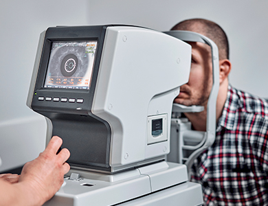
What to Expect During Your Retinal Vein Occlusion Evaluation
Our comprehensive assessment includes:
Dilated fundus examination
High-resolution optical coherence tomography (OCT) to detect and quantify macular edema
Fundus photography and, when indicated, fluorescein angiography to evaluate blood flow and leakage
The evaluation is non-invasive and painless (aside from mild stinging from dilating drops). We also perform visual acuity testing and discuss your overall health history.
Treatment Options
While the blockage itself cannot be reversed, modern treatments effectively manage complications and improve or stabilize vision:
Intravitreal anti-VEGF injections (most common first-line treatment for macular edema)
Intravitreal steroid implants
Laser photocoagulation (focal or panretinal) for persistent edema or neovascularization
In rare advanced cases, vitreoretinal surgery
Close monitoring allows us to tailor therapy and prevent secondary issues like neovascular glaucoma.
If you have experienced sudden vision changes or have risk factors for retinal vein occlusion, schedule an urgent evaluation with a retina specialist. Early intervention can significantly protect your sight. Contact our team today for more information or to book your comprehensive retinal assessment.

Contact Us Let’s keep in touch!
Office Hours
- Monday 8:00am - 5:00pm
- Tuesday 8:00am - 5:00pm
- Wednesday 8:00am - 5:00pm
- Thursday 8:00am - 5:00pm
- Friday 8:00am - 5:00pm
- Saturday Closed
- Sunday Closed
Closed for lunch from 12 pm-1 pm
© 2026 Four States Retina | All rights Reserved | Accessibility Statement | Privacy Policy | Sitemap
Powered by: