
Posterior Vitreous Detachment
Posterior Vitreous Detachment (PVD): Understanding This Common Age-Related Change and Protecting Your Retina
As retina specialists, we frequently diagnose posterior vitreous detachment (PVD), one of the most common events in the aging eye. PVD occurs when the vitreous gel—the clear, jelly-like substance that fills the eye—naturally shrinks and separates from the retina. This process is a normal part of aging, typically beginning after age 50, and affects the majority of people by their 70s or 80s.
While PVD itself is usually benign, the separation can occasionally cause a retinal tear in 10–15% of cases. An untreated tear can progress to retinal detachment, a serious condition that threatens permanent vision loss. Prompt evaluation by a retina specialist is essential when symptoms appear suddenly.
What Is Posterior Vitreous Detachment?
The vitreous is attached to the retina in youth. With age, it liquefies (syneresis) and condenses, eventually pulling away from the retinal surface. This separation often produces a ring-shaped floater known as a Weiss ring—a classic sign of complete PVD.
What Symptoms Should You Watch For?
Patients often describe:
Sudden appearance of numerous new floaters (specks, threads, or clouds drifting across vision)
Flashes of light (arcs, streaks, or lightning-like flashes, especially in dim light or peripheral vision)
When Is It an Emergency?
Seek immediate care from a retina specialist if you notice:
A sudden shower of new floaters
New or persistent flashes
A shadow, curtain, or dark veil moving across your vision
Blurred or decreased vision
These may signal a retinal tear or early detachment.
What to Expect During Your PVD Evaluation
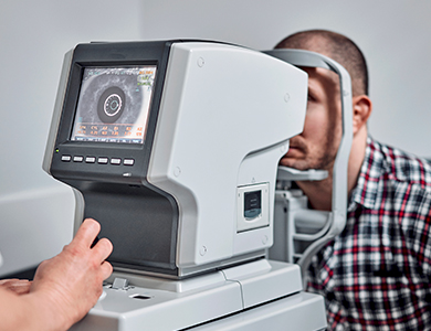
We perform a thorough dilated retinal examination, often the same day or next day for acute symptoms. This includes:
Detailed peripheral retinal exam using indirect ophthalmoscopy
High-resolution optical coherence tomography (OCT) to visualize the vitreoretinal interface
Management and Treatment
Uncomplicated PVD: Observation, patient education, and follow-up as needed
Retinal tear: In-office laser retinopexy to seal the tear and prevent progression to detachment
Retinal detachment (if present): Surgical intervention (vitrectomy, scleral buckle, etc.)
If you are experiencing new floaters, flashes, or any sudden change in vision, do not delay—contact a retina specialist immediately. Most serious complications are preventable with early detection and treatment. Our team is here to provide urgent evaluation and personalized care.

Contact Us Let’s keep in touch!
Office Hours
- Monday 8:00am - 5:00pm
- Tuesday 8:00am - 5:00pm
- Wednesday 8:00am - 5:00pm
- Thursday 8:00am - 5:00pm
- Friday 8:00am - 5:00pm
- Saturday Closed
- Sunday Closed
Closed for lunch from 12 pm-1 pm
© 2026 Four States Retina | All rights Reserved | Accessibility Statement | Privacy Policy | Sitemap
Powered by: