
Flashes & Floaters
Flashes and Floaters: Urgent Retinal Evaluation to Protect Your Vision
These common visual symptoms are often the first warning signs of potentially serious retinal conditions, including posterior vitreous detachment (PVD), retinal tears, or even retinal detachment. While many cases are benign, sudden onset or a sudden increase in symptoms requires prompt assessment to prevent permanent vision loss.
Early intervention by a retina specialist can make the critical difference between a simple observation and the need for laser treatment or surgery.
What Are Flashes and Floaters?
Floaters appear as small specks, threads, cobwebs, or clouds drifting across your field of vision. They are caused by tiny clumps of gel or cells inside the vitreous—the clear, jelly-like substance that fills the eye.
Flashes of light (often described as lightning streaks or arcs, especially in peripheral vision) occur when the vitreous tugs on the retina during separation.
What Causes These Symptoms?
The most common cause in adults over 50 is posterior vitreous detachment (PVD), a natural aging process where the vitreous gel shrinks and pulls away from the retina. This often produces a ring-shaped floater called a Weiss ring.
However, in about 10–15% of PVD cases, the traction can cause a retinal tear. If untreated, fluid can seep under the tear, leading to retinal detachment—a sight-threatening emergency.
When Should You Seek Immediate Care?
Contact a retina specialist urgently if you experience:
Sudden onset of numerous new floaters
New or persistent flashes of light
A shadow, curtain, or veil progressing across your vision
Blurred or decreased vision
These can indicate a retinal tear or detachment requiring prompt intervention.
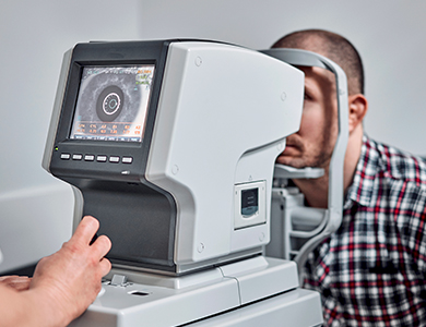
What to Expect During Your Urgent Retinal Evaluation
We perform a comprehensive dilated retinal examination, including:
Dilated fundus exam to visualize the peripheral retina
High-resolution optical coherence tomography (OCT) to assess the vitreoretinal interface
B-scan ultrasonography if vitreous hemorrhage obscures the view
The exam is non-invasive and painless (aside from temporary blurring from dilating drops). Most patients are seen the same day or next day when symptoms are acute.
Treatment Options
Benign PVD: Observation and education
Retinal tear: In-office laser retinopexy to seal the tear and prevent detachment
Retinal detachment: Surgical repair (vitrectomy, scleral buckle, or pneumatic retinopexy)
If you are experiencing new or worsening flashes and floaters, do not wait—schedule an urgent evaluation with a retina specialist. Many serious complications are preventable with timely care. Contact our team today for immediate assistance.

Contact Us Let’s keep in touch!
Office Hours
- Monday 8:00am - 5:00pm
- Tuesday 8:00am - 5:00pm
- Wednesday 8:00am - 5:00pm
- Thursday 8:00am - 5:00pm
- Friday 8:00am - 5:00pm
- Saturday Closed
- Sunday Closed
Closed for lunch from 12 pm-1 pm
© 2026 Four States Retina | All rights Reserved | Accessibility Statement | Privacy Policy | Sitemap
Powered by: