
Macular Hole
Macular Hole: Advanced Diagnosis and Surgical Repair to Restore Central Vision
Macular hole is a condition in which a full-thickness defect develops in the center of the macula—the part of the retina responsible for sharp, detailed central vision. Untreated macular holes almost always lead to significant and permanent central vision loss. Fortunately, modern vitreoretinal surgery offers excellent success rates, with many patients regaining functional central vision when treated early.
Macular holes most commonly occur in people over 60, particularly women, and are often related to age-related changes in the vitreous gel.
What Is a Macular Hole?
Macular holes typically form during posterior vitreous detachment (PVD) when the vitreous gel pulls abnormally on the fovea (the very center of the macula), creating traction that tears the retinal tissue.
Symptoms of Macular Hole
Patients usually notice symptoms in one eye:
Blurred or distorted central vision
A dark or missing spot in the center of vision (central scotoma)
Metamorphopsia (straight lines appear wavy or bent)
Difficulty reading, recognizing faces, or performing detailed tasks
What to Expect During Your Macular Hole Evaluation
We perform a comprehensive assessment, including:
Dilated fundus examination to visualize the hole
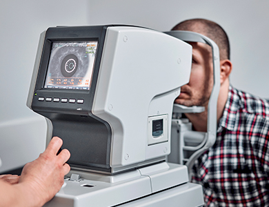
High-resolution optical coherence tomography (OCT)—the gold standard for staging, measuring hole size, and assessing surrounding retinal changes
Treatment: Vitrectomy Surgery with Membrane Peeling
The standard treatment for full-thickness macular holes is pars plana vitrectomy combined with internal limiting membrane (ILM) peeling and gas tamponade. This outpatient procedure involves:
Removing the vitreous gel
Carefully peeling the internal limiting membrane to relieve traction
Placing a gas bubble to press the retina flat and promote hole closure
When to Seek Care
If you notice a sudden or progressive central blur, dark spot, or distorted vision in one eye, schedule an urgent evaluation with a retina specialist. Early diagnosis and surgery (ideally within weeks to months of symptom onset) provide the best chance for visual recovery.
Contact our team today for prompt assessment and expert surgical care.

Contact Us Let’s keep in touch!
Office Hours
- Monday 8:00am - 5:00pm
- Tuesday 8:00am - 5:00pm
- Wednesday 8:00am - 5:00pm
- Thursday 8:00am - 5:00pm
- Friday 8:00am - 5:00pm
- Saturday Closed
- Sunday Closed
Closed for lunch from 12 pm-1 pm
© 2026 Four States Retina | All rights Reserved | Accessibility Statement | Privacy Policy | Sitemap
Powered by: